A New Perspective on PRP Therapy
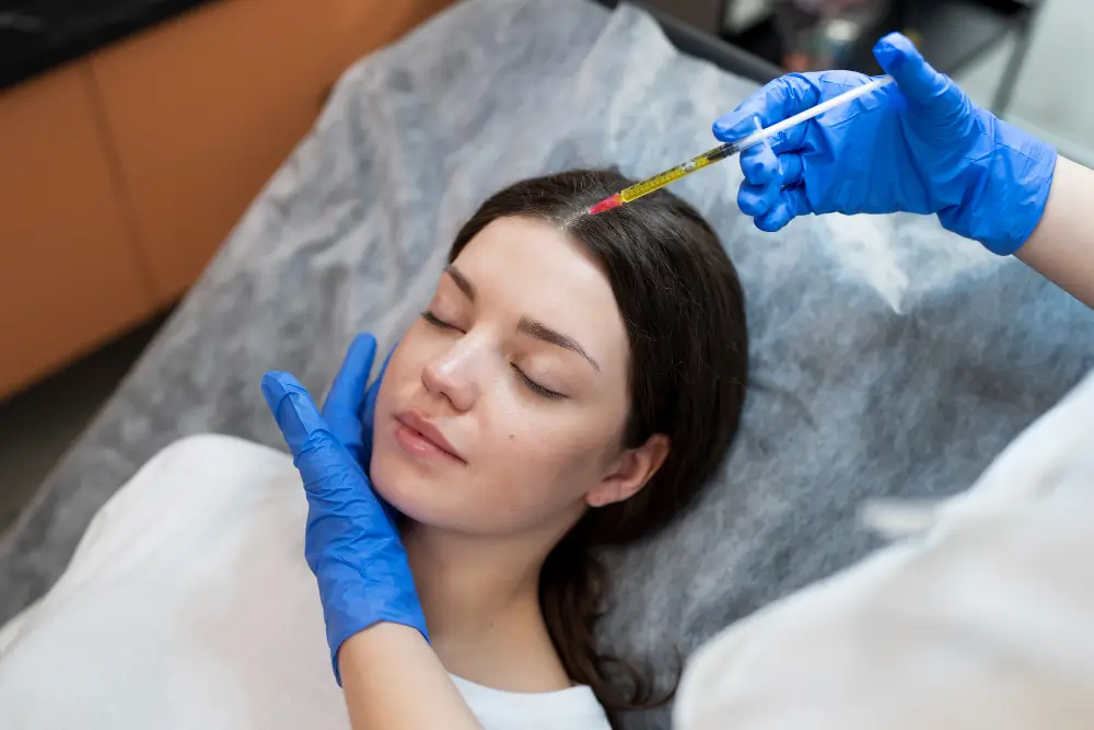
Platelet-Rich Plasma (PRP) has become one of the most widely used regenerative therapies in modern orthobiologics. For years, clinicians have focused on two primary measures of PRP quality: platelet concentration and total platelet dose.
These parameters remain important because platelets serve as the primary reservoir of growth factors and signaling molecules involved in tissue repair.
Yet as the use of PRP has expanded, an important observation has emerged. Two PRP preparations with nearly identical platelet counts can produce very different clinical outcomes. This variability has led researchers to examine factors beyond simple platelet concentration. Current evidence suggests that platelet number alone may not fully explain the biological activity of a PRP preparation. The functional quality of the platelets, the integrity of their growth factor cargo, the methods used during processing, and the patient’s own biology all appear to influence the final therapeutic effect.
As a result, the field is gradually shifting from a purely quantitative model toward a more comprehensive understanding of PRP as a dynamic biologic system.
Why Platelets Matter
These stem cell therapy options encourage the “proliferation”, growth, and repair of ligament, joint, and musculoskeletal tissues. This treatment involves the injection of a high-concentration dextrose (sugar water) solution into the targeted area. The presence of the solution in the affected area causes a mild irritation to the tissue leading to increased blood flow and nutrients to the area which then signal the body to heal the damaged tissue.
Prolozone is the addition of ozone gas to the prolotherapy solution. The additional ozone combines with signaling molecules to act as regulators of the immune system to lessen inflammation. This results in increased function as well as pain reduction.
Balance Matters
Regenerative signaling relies on appropriate relationships among growth factors and cytokines. Excessive or imbalanced signaling may promote fibrosis or dysregulated healing rather than tissue regeneration.
Timing Matters
The release pattern of growth factors affects how cells respond. Different phases of healing require different signals, making release kinetics an important consideration.
More Is Not Always Better
Higher concentrations do not necessarily translate into better outcomes. Biological systems frequently demonstrate saturation effects, and excessive stimulation can sometimes produce diminishing returns.
For these reasons, PRP efficacy appears to depend on more than platelet count alone. Platelet integrity, activation status, growth factor composition, leukocyte content, and patient-specific variables all contribute to the final biologic effect.
Next-Generation Orthobiologics
Traditional PRP systems were designed primarily to separate and concentrate platelets from whole blood. While effective, these methods generally conclude once the platelet-rich fraction has been isolated.
Emerging technologies seek to further refine the biologic product before reinjection through additional processing steps intended to optimize signaling, reduce unwanted components, and concentrate beneficial proteins.
These approaches generally involve three major stages:
- Platelet activation
- Filtration
- Plasma protein concentration
Step 1: Platelet Activation
Activation determines when and how platelets release their stored growth factors.
A variety of activation methods have been developed, including calcium chloride activation, thrombin activation, freeze-thaw platelet lysates, collagen-based activation, and surface-mediated activation systems. Each approach influences the timing and magnitude of growth factor release.
One particularly interesting strategy involves activation through contact with specialized glass surfaces. When platelets encounter negatively charged glass, physiologic activation pathways are triggered, leading to platelet adhesion, aggregation, and degranulation. This process stimulates the release of important regenerative mediators including PDGF, TGF-β, and VEGF.
Glass bead technology has also been used in autologous cytokine therapies such as Orthokine® and Autologous Conditioned Serum (ACS) systems. In these systems, blood is incubated in syringes containing borosilicate glass beads, which stimulate monocytes and other immune cells through surface contact.
Research has demonstrated increased production of anti-inflammatory mediators such as interleukin-1 receptor antagonist (IL-1Ra), along with elevations in IL-10 and multiple regenerative growth factors. While some pro-inflammatory cytokines may also increase during incubation, the overall cytokine profile tends to favor an anti-inflammatory environment.
In addition to growth factor release, activation systems may contribute to the formation of biologic scaffolds that influence the spatial and temporal distribution of regenerative signals after injection.
Step 2: Filtration and Product Refinement
Filtration serves as an additional processing step designed to improve the consistency of the final orthobiologic product.
Potential benefits include:
- Reduction of cellular debris
- Removal of unwanted particulate material
- Improved injectability
- Enhanced product uniformity
- Reduction of certain inflammatory cellular components
When activation systems involving glass surfaces are used, filtration can also help remove residual particulate material before administration.
Although filtration is not intended to sterilize the product, it can help create a cleaner and more controlled biologic environment by refining the cellular and physical characteristics of the final preparation.
Step 3: Plasma Protein Concentration
One of the most significant developments in orthobiologics involves the concentration of beneficial plasma proteins from platelet-poor plasma (PPP).
Historically, PPP was often considered a secondary byproduct of PRP preparation. Newer technologies suggest that this fraction may contain important therapeutic molecules that deserve greater attention.
Modern ultrafiltration systems utilize polysulfone hollow-fiber membranes to selectively remove plasma water while retaining larger proteins and biologically active molecules. This process can concentrate alpha-2 macroglobulin (A2M), insulin-like growth factor-1 (IGF-1), hepatocyte growth factor (HGF), fibrinogen, cytokines, and additional regulatory proteins.
Recent investigations have demonstrated substantial enrichment of A2M and IGF-1 through these ultrafiltration methods.
A2M is of particular interest because it functions as a broad-spectrum inhibitor of cartilage-degrading enzymes. Research suggests that A2M may help slow cartilage breakdown and reduce protease-mediated joint degeneration associated with osteoarthritis.
This represents a potentially important shift in thinking. Rather than viewing biologic therapies solely as regenerative stimulants, plasma-derived concentrates may also provide protective and anti-catabolic effects that help preserve tissue integrity.
By concentrating beneficial proteins while reducing excess plasma volume, these technologies create a biologically enriched product that may complement traditional PRP treatments.
What This Means for Patients
For patients considering PRP or other orthobiologic treatments, these advances highlight an important reality: not all PRP preparations are necessarily identical.
Historically, discussions about PRP quality have focused on platelet concentration alone. While platelet dose remains an important factor, emerging research suggests that the biological activity of a PRP preparation may also depend on how the product is processed, activated, filtered, and delivered.
The quality of the platelets, the composition of growth factors and cytokines, and the presence of supportive plasma proteins may all contribute to the overall therapeutic effect.
This does not mean that older PRP systems are ineffective. Rather, it reflects the ongoing evolution of regenerative medicine as clinicians and researchers work to better understand the complex biological mechanisms involved in tissue healing.
Patients should recognize that individual outcomes depend on many factors, including:
- The severity and chronicity of the condition being treated
- Age and overall health status
- Metabolic health and inflammatory burden
- The specific PRP or orthobiologic preparation used
- The experience and technique of the treating clinician
- Rehabilitation and post-procedure compliance
Modern orthobiologic approaches are increasingly focused on creating a more favorable healing environment rather than simply delivering a higher number of platelets. By optimizing growth factor signaling, reducing unwanted inflammatory components, and concentrating beneficial plasma proteins, newer preparation methods seek to support the body’s natural repair processes in a more targeted and biologically sophisticated manner.
As research continues to evolve, the future of regenerative medicine may be defined not only by how much biology is delivered, but by how effectively those biological signals are organized and directed toward healing.
The Future of PRP: A More Complete Biological Model
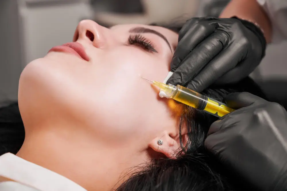
Platelet concentration remains an important metric in regenerative medicine. Without adequate platelet numbers, sufficient growth factor delivery becomes difficult to achieve.
However, growing evidence suggests that successful outcomes depend on much more than platelet count alone. Factors such as platelet functional integrity, growth factor composition, activation methods, processing techniques, cytokine balance, plasma protein enrichment, patient age, metabolic health, oxidative stress, and systemic inflammation may all influence the biological behavior of PRP.
The next phase of orthobiologic development is likely to focus on understanding and optimizing these interconnected variables rather than simply increasing platelet concentration.
PRP should increasingly be viewed as a sophisticated biologic ecosystem rather than a product defined by a single laboratory measurement.
As regenerative medicine continues to evolve, the emphasis will likely shift from maximizing platelet quantity toward preserving and directing the biological signals that ultimately drive healing. In this emerging framework, clinical success depends not only on how many platelets are delivered, but on how effectively the entire regenerative environment is engineered to support tissue repair and recovery.
References
- Costa, F. R., Purita, J., Martins, R., Pires, L., Mahmood, A., Santos, G. S., Kruel, A., Protásio Netto, J., & Lana, J. F. (2026). Beyond Platelet Count: Rethinking Platelet-Rich Plasma Efficacy Through Growth Factor Biology and Functional Quality. Life, 16(2), 188. https://doi.org/10.3390/life16020188
- Edwards, D. R., Murphy, G., Reynolds, J. J., Whitham, S. E., Docherty, A. J. P., Angel, P., & Heath, J. K. (1987). Transforming growth factor beta modulates the expression of collagenase and metalloproteinase inhibitor. The EMBO Journal, 6(7), 1899–1904.
- Morris C. D. (1968). Observations on the effect of glass beads on platelet aggregation and its relation to platelet stickiness. Thrombosis et diathesis haemorrhagica, 20(3), 345–353.
- Wang, S., Wei, X., Zhou, J., Zhang, J., Li, K., Chen, Q., Terek, R., Fleming, B. C., Goldring, M. B., Ehrlich, M. G., Zhang, G., & Wei, L. (2014). Identification of α2-macroglobulin as a master inhibitor of cartilage-degrading factors that attenuates the progression of posttraumatic osteoarthritis. Arthritis & rheumatology (Hoboken, N.J.), 66(7), 1843–1853. https://doi.org/10.1002/ art.38576
- Meijer H, Reinecke J, Becker C, Tholen G, Wehling P. The production of anti-inflammatory cytokines in whole blood by physico-chemical induction. Inflamm Res. 2003;52(10):404-407. doi:10.1007/s00011-003-1197-1
- Baltzer AW, Moser C, Jansen SA, Krauspe R. Autologous conditioned serum (Orthokine) is an effective treatment for knee osteoarthritis. Osteoarthritis Cartilage. 2009;17(2):152-160. doi:10.1016/j.joca.2008.05.010
- Wehling P, Moser C, Frisbie D, et al. Autologous conditioned serum in the treatment of orthopedic diseases: the Orthokine therapy. BioDrugs. 2007;21(5):323-332.
- Magalon J, Bausset O, Veran J, et al. Physico-chemical factors influencing autologous conditioned serum purification. Blood Transfus. 2014;12(Suppl 1):s573-s584. doi:10.2450/2013.0250-12
- Rutgers M, Saris DBF, Dhert WJA, Creemers LB. Cytokine profile of autologous conditioned serum for treatment of osteoarthritis, in vitro effects on cartilage metabolism and intra-articular levels after injection. Arthritis Res Ther. 2010;12(3):R114. doi:10.1186/ar3050
- Fernández-Pernas P, Barrachina L, Marquina M, et al. A comparative review of autologous conditioned serum and autologous protein solution for treatment of osteoarthritis in horses. Front Vet Sci. 2021;8:602978. doi:10.3389/fvets.2021.602978
- Pishgahi A, Roshangar L, Afkham-Daghdaghan M, Pourabbas B, Yousefi M. The therapeutic effects of autologous conditioned serum on knee osteoarthritis: an animal model. BMC Res Notes. 2022;15:280. doi:10.1186/ s13104-022-06166-1
- Knieć K, Kowalski K, Domżalski M, Gajewski M. Deep or superficial? Autologous conditioned serum for cervical pain with degenerative disc disease — a randomized controlled trial. Pain Res Manag. 2025. doi:10.1155/prm/6691691
- Showel, K. K., Evans, C. H., De la Vega, R. E., Hawse, G. P., Gonzalez Carta, K., Saris, D. B. F., Sellon, J. L., & Boettcher, B. J. (2025). Preparation of alpha-2 macroglobulin–rich plasma from human blood—technique and compositional analysis of plasma, platelet-rich plasma, platelet-poor plasma, and alpha-2 macroglobulin–rich plasma. Journal of Cartilage and Joint Preservation, 5(4), Article 100255. https://doi.org/10.1016/j.jcjp.2025.100255
- Everts P, Onishi K, Jayaram P, Lana JF, Mautner K. Profound properties of protein-rich, platelet-rich plasma matrices as novel, multi-purpose biological platforms in tissue repair, regeneration, and wound healing. Int J Mol Sci. 2024;25(14):7914. doi:10.3390/ijms25147914
- Vandooren J, Itoh Y. Alpha-2-macroglobulin in inflammation, immunity and infections. Front Immunol. 2021;12:803244. doi:10.3389/fimmu.2021.803244
Contact Us
Contact your physician at Hawaii Island Stem Cell Therapy and Regenerative Medicine to schedule an appointment to find out if these therapies are right for you.
